
Dr. (Prof) Suvro Banerjee
Senior Cardiac Doctor in Kolkata
MD, MRCP (UK), FRCP (EDIN), FRCP (London), FICC, FCSI, FESC, FACC, FSCAI,
Senior Consultant Interventional Cardiologist, Apollo Multispeciality Hospitals, Kolkata
Senior Cardiac Doctor in Kolkata
MD, MRCP (UK), FRCP (EDIN), FRCP (London), FICC, FCSI, FESC, FACC, FSCAI,
Senior Consultant Interventional Cardiologist, Apollo Multispeciality Hospitals, Kolkata
HOW HEART WORKS :
The muscles and various body organs (such as lungs, kidneys and brain) need nourishment in order to function efficiently. This nourishment is provided by blood rich in oxygen. Circulation of blood is essential for oxygen to reach the muscles and organs. The main function of the heart is to pump blood to maintain an efficient circulation.
The function of the heart therefore can be compared to a domestic pump used for lifting water. A pump comprises of a mechanical part (machine) which produces the force needed for lifting water, and an electrical circuit that runs the machine. The water, being driven by the pump, flows through pipes which are provided with valves to ensure unidirectional flow of water.
The heart has four 'chambers'. The two upper chambers called left and right 'atrium' and two lower chambers called left and right 'ventricles'. These chambers are walled by powerful muscles which pump blood to maintain the circulation.
Pure oxygen rich blood flows from the left sided heart chambers into a large artery known as aorta through which it reaches the different organs of the body. The oxygen is extracted from the blood by these body organs and in turn waste products like carbon dioxide are released back into the blood stream. Impure blood from the different body organs reaches the right sided chambers through channels known as systemic veins. The heart then pumps this blood into the lungs through a larger channel called pulmonary artery. The blood is purified in the lungs and returned to the left side of the heart through another set of channels called pulmonary veins. The movement of the blood through the heart is regulated by a system of valves, which ensure that the blood flows in the correct direction.
A heart beat results when the chambers contract to pump the blood. Just as a pump requires electricity for it to run, the heart has an inherent electric circuit that ensures constant pumping of the heart muscles.
Besides supplying blood to other organs, the heart itself needs nutrition which is provided by special arteries known as coronary arteries.
Knowledge of the anatomy and the function of the heart will help us to understand the cause and effect of different kinds of heart diseases.
COMMON HEART DISEASES :
The heart however is not always as romantic as it sounds. Each component of the heart has the potential for misbehaviour causing significant illness or even death.
ISCHAEMIC HEART DISEASE:
This occurs when there is narrowing of the coronary arteries (which supply nutrition to the heart). Narrowing occurs due to deposition of cholesterol and other substances in the wall of the arteries. This may cause chest pain (angina) on exertion.
MYOCARDIAL INFARCTION (HEART ATTACK):
This occurs when there is complete obstruction to flow of blood in the coronary artery. The segment of the heart muscle supplied by the artery may be permanently damaged.
HEART FAILURE:
This occurs because of the inability of the heart to pump blood effectively resulting in poor nourishment of body organs. Causes are many including high blood pressure, anaemia and diseases of coronary arteries, heart muscles or heart valves.
CARDIAC DYSRHYTHMIA:
Usually results from disease of the electrical circuit of the heart. There may be slowing of heart rate (bradyarrythmia or heart block) or abnormally high heart rate (tachyarrythmia). Palpitation, dizziness or black-out may result.
VALVE DISEASE:
Disease of valve may result from rheumatic fever, birth defects involving the valve, degeneration of the valve as part of aging process or valve infection. The valve may become narrow or leaking or both.
ENDOCARDITIS:
Infection of heart valve.
CONGENITAL HEART DISEASE:
Defect is present since birth, although the manifestation may occur after a variable time interval.
SEPTAL DEFECTS:
Usually congenital. Holes in the walls that partition the heart chambers.
CARDIOMYOPATHY:
Disease of heart muscles which may cause heart failure.
PERICARDIAL DISEASES:
Disease of the sac that surrounds the heart. There may be inflammation of the sac (pericarditis), fluid collection inside the sac (pericardial effusion) or thickening of the sac (constrictive pericarditis).
SYMPTOMS OF HEART DISEASES :
ISCHAEMIC HEART DISEASE:
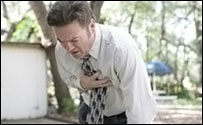
This is a common symptom of ischaemic heart disease. The pain usually occurs centrally in the chest. It may pass down to left arm, to the jaw or to upper part of the tummy. Occasionally it may radiate to right shoulder, to the back or sometimes may not radiate at all. It is variously described, sometimes as a 'crushing pain' or may simply be 'a sense of heaviness over the chest'. Usually it comes with exertion and is relieved with rest. In severe cases, it may start even during period of inactivity.
It is important to remember that chest pain is not always due to some heart ailments. Other causes such as indigestion, chest infection, inflammation of muscles and joints of the rib cage may also cause chest pain. Occasionally heart attack may occur even without chest pain, particularly in elderly and in diabetics.
SHORTNESS OF BREATH :

This usually occurs with exertion, although in more severe cases it may be present even at rest. This may be associated with 'orthopnoea', where breathlessness is aggravated on lying flat or 'paroxysmal nocturnal dyspnoea', where sudden bout of breathlessness may wake you up from sleep leaving you gasping for breath. Shortness of breath may indicate heart failure, however lung disease, obesity, anaemia or even physical deconditioning may cause breathlessness on exertion.
PALPITATION :
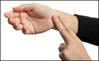
This is awareness of one's own heartbeat. It may be heightened awareness of normal heartbeat or due to abnormal beating of the heart. Palpitation may also occur due to anxiety, mental stress or excitement.
DIZZINESS :
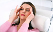
'Light-headedness' or 'feeling as if may fall or faint', may be due to abnormal heart rhythm or abnormal blood pressure. It may also be due to disorders other than heart disease.
BLACK OUT :
Sudden loss of consciousness is usually an ominous sign. Disorders of heart rhythm may cause black out. Other diseases such as epilepsy or stroke may also have similar manifestation.
SWELLING OF FEET :

This may have different causes. Heart failure is one of them. Others include kidney disease, liver disease, anaemia, low protein in blood, diseases of venous or lymphatic channels of the leg.
Even in absence of symptoms, if you have multiple risk factors for coronary artery disease, such as high blood pressure, diabetes, high cholesterol, history of heart disease in your family or habit of smoking then also you should consider seeing a doctor for advice.
It is important to remember that occasionally early heart disease may not be associated with any symptom and the disease may be detected incidentally during routine examination.
CARDIAC INVESTIGATIONS :
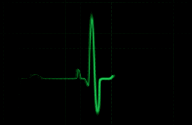
ELECTROCARDIOGRAM (ECG):
An ECG is a graphic recording of the electrical activity of the heart. It is primarily used as a screening test for patients with cardiac symptoms.
TREADMILL TEST (TMT):
Also called exercise stress test or exercise tolerance test. This test can detect coronary artery disease even when the ECG is normal. The test is advocated in patients with chest pain to exclude significant coronary artery disease.
HOLTER MONITOR ( 24-HOUR AMBULATORY ECG MONITOR):
This is a continuous recording of your heart's electrical activity, made by a small light-weight recorder strapped to your body that operates whilst you carry on with your daily routine. The test may be necessary if you complain of symptoms such as palpitation or blackouts or in some cases where your ECG is abnormal.
24-HOUR BLOOD PRESSURE MONITOR:
This is similar to Holter monitor, except that your blood pressure is measured over a continuous period instead of your heart's electrical activity. The blood pressure that is measured in your doctor's chamber may be unusually high for you and may not reflect your usual blood pressure. This phenomenon (called White Coat Hypertension) may cause inappropriate treatment of your blood pressure. The test identifies those with white-coat hypertension and helps in the assessment of the efficacy of drug therapy.
IMPLANTABLE LOOP RECORDER:
A small device is implanted under the skin which continuously records the ECG. It can provide vital clues in cases of unexplained symptoms such as palpitation or blackout. It could be left for more than a year if necessary and therefore specially useful where symptoms are infrequent.
SIGNAL-AVERAGED ECG:
This test identifies patients who are at risk for developing potentially lethal heart rhythms.
HEART RATE VARIABILITY (HRV):
In normal healthy individuals heart rate varies significantly over 24 hours. Reduced heart rate variability is associated with increased cardiovascular morbidity and mortality.
ECHOCARDIOGRAPHY & COLOUR DOPPLER (TTE):
This test utilises ultrasound to obtain still and moving images of the beating heart. It provides information on the size, structure and function of the heart chambers, motion of the heart valves and assessment of blood flow through the heart and the vessels. It is a painless procedure and free from radiation exposure.
TRANSOESOPHAGEAL ECHOCARDIOGRAPHY (TEE):
A probe with an ultrasound device at its tip is passed into the patient's gullet to image the heart from behind. It usually provides better image resolution and can also visualise the posterior structures of the heart that cannot be seen well by transthoracic echocardiography (TTE). It is also used in the operating theatre especially in cardiac surgery.
STRESS ECHOCARDIOGRAPHY:
Intravenous medication is used to stimulate the heart. This unmasks latent coronary disease, which shows up as abnormal contraction of heart muscles. The test is particularly useful for patients who are not fit for exercise. The test is painless and non-invasive.
CARDIAC INTERVENTIONS :
CARDIAC CATHETERISATION AND CORONARY ANGIOGRAPHY:
This is a diagnostic procedure. Long, slender, flexible tubes called catheters are inserted usually through the groin and passed through the vessels into the heart chambers and into the arteries supplying the heart (coronary arteries). Pressures inside the heart chambers are measured and a dye is injected to visualise the coronary arteries for any evidence of narrowing.
CORONARY ANGIOPLASTY WITH STENTING:
Angina and heart attack result from narrowing or occlusion of coronary arteries. In selected cases, passing a balloon through the artery may open up a narrowed coronary artery. Deployment of an artificial mesh (stent) helps to keep the artery open.
PERIPHERAL ANGIOPLASTY:
Balloon catheters passed through skin can open up narrowed arteries in the limbs, neck or kidneys. Lasting benefit is possible in majority of cases.
PULMONARY AND MITRAL BALLOON VALVULOPLASTY:
A narrowed heart valve is opened up by the passage of a balloon passed through the skin under local anaesthesia, thereby reducing the risk and morbidity of a surgical procedure. Outcome is very similar to surgery.
SINGLE AND DUAL CHAMBER PERMANENT PACEMAKER IMPLANTATION:
Implantation of pacemaker becomes necessary where there is abnormality in the electrical activity of the heart resulting in heart block or abnormal heart rhythm. Depending on the type of abnormality, a single or dual chamber pacemaker may be prescribed.
ELECTROPHYSIOLOGY STUDY (EPS):
This is a diagnostic test that helps management of patients with abnormal heart rhythm. Thin, long catheters are inserted through neck or groin into the heart with a view to reproduce the rhythm disturbances, locate the site of abnormal rhythm, predict the efficacy of drug therapy and plan further management.
RADIOFREQUENCY CATHETER ABLATION:
An almost painless procedure which can cure electrical short circuits in the heart. Useful in selected patients with abnormally fast heart rhythms.
ICD:
It is a device which is useful in patients with history of or high risk of sudden cardiac arrest. It is implanted just like a pacemaker. It constantly analyses the heart rhythm and in case of cardiac arrest it reverts the heart rhythm to normal by delivering an electric shock. Moreover, it also provides back-up pacemaker function if the heart rate drops.
CARDIAC RESYNCHRONISATION THERAPY (CRT / BIVENTRICULAR PACEMAKER):
It is a special variety of pacemaker which is useful in patients with heart failure who are not responding to medical treatment. It improves the pumping efficiency of the heart. Not only it improves symptoms but also prognosis in selected cases.